√無料でダウンロード! sinus rhythm with 1st degree av block right bundle branch block 170983-Sinus rhythm with 1st degree av block right bundle branch block
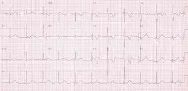
Dec 05, · Right Bundle Branch Block (RBBB) – the basics Right Bundle Branch Block (RBBB) with 1st degree AV block on a full ECG In many people, this does not cause abnormalities of the ECG It often indicates right sided heart disease In the normal heart, the depolarisation of the septum occurs from right to leftJun 07, 16 · Wiring of heart Sinus rhythm means that the electrical activity that controls the contraction of the heart, starts in the sinus node Right bundle branch block means that the right bundle (part of the electrical wiring) does not conduct the impulse which changes the appearance of the ekg Right bundle branch block as opposed to left bundle branch block is usually benignJan 31, 10 · Sinus bradycardia with first degree AV block Sinus bradycardia is evident from the long RR interval of 1280 ms, corresponding to a heart rate of 47 per minute PR interval is also prolonged at about 3 msec The combination can occur in vagotonic states or in those on beta blockers or other drugs which suppress both the sinus node and the AV

Evaluation And Treatment Of Av Block And Intraventricular Conduction Disturbances The Cardiology Advisor
Sinus rhythm with 1st degree av block right bundle branch block
Sinus rhythm with 1st degree av block right bundle branch block-Oct 02, · A PR interval > 0 ms suggests a firstdegree AV block Firstdegree AV block When PR interval > 0 ms, each P wave is followed by a QRS complex Seconddegree AV block (Mobitz I or Wenckelbach) The PR interval steadily increases until the impulse transmission fails (skipped heartbeat, and missing QRS complex)In persons with or without overt heart disease, LBBB is associated with a higher risk of mortality and morbidity from myocardial infarction, heart failure, and arrhythmias such as highgrade AV block 17 (Fig 323) Right bundle branch block (RBBB) The common diagnostic criteria for RBBB are listed in Table 311 In contrast to LBBB, RBBB


3rd Degree Av Block Ecg 1 Learntheheart Com
Sinus Rhythm means you heart is beating at a steady consistent rate A first degree AV block means that the electrical signal that starts in the Atria (upper chambers) of the heart and is relayed to the Ventricles (lower chambers) of the heart, is taking too long to get there It's sometimes referred to on the EKG as a prolonged PR timeSeconddegree atrioventricular block (AV block) is a disease of the electrical conduction system of the heartIt is a conduction block between the atria and ventriclesThe presence of seconddegree AV block is diagnosed when one or more (but not all) of the atrial impulses fail to conduct to the ventricles due to impaired conductionFeb 13, 16 · Although the combination of right bundle branch block, left anterior or posterior fascicular block, and first degree AV block is often called "trifascicular block" the term in this context is a bit of a misnomer First degree AV block is not a true block because there is a 11 relationship between Pwaves and QRS complexes
Heart block happens when the flow of electricity from the top to bottom of the heart is delayed or blocked at some point along the pathway There are three main types of heart block AV (atrioventricular) heart blocks Bundle branch blocks Tachybrady syndromeDec 29, 15 · A first degree AVblock is said to be present when the PRinterval is greater than 0 ms in the setting of a sinus rhythm Figure 2 Sinus rhythm with first degree AVblock in a 16yo M s/p seizure The prolonged PRinterval is caused by a delay in conduction, usually within the AVnode but sometimes slightly above it in the atrial tissue orCHRONIC bifascicular bundle branch block or left bundle branch block (LBBB) can progress to complete heart block (CHB) with the risk of sudden perioperative death 1–3 In the perioperative period, different factors can induce a prolongation of atrioventricular (AV) conduction and increase the risk for bradyarrhythmias
Jul 07, · The rhythm strip shows sinus rhythm with two QRS configurations;Note that the first three account for almost 90% of EKG tracings with leftaxis deviation Normal variation elderly or obese patients Left ventricular hypertrophy Left anterior fascicular block Left bundle branch block Inferior wall myocardial infarction Ventricular ectopic rhythms (eg, ventricular tachycardia)Mar 02, 15 · In addition i) the PR interval of conducting beats is long ( ~ 024 second) — with 1stDegree AV Block being more common with Mobitz I rather than Mobitz II;


Pin On Ekg


Pin On Ekg
Aug 01, 1999 · sinus rhythm;Oct 01, 10 · Two days after his presentation, ECG showed a sinus rhythm with 1st degree AV block, right bundle branch block (RBBB) and a left anterior fascicular block Echocardiogram was performed and showed normal left ventricular size with ejection fraction of 68%, dilated right ventricle with a right ventricular systolic pressure was 53 mmHgRight bundle branch block (RBBB—see figure Right bundle branch block) can occur in people with no evidence of heart disease It may also occur with anterior myocardial infarction, indicating substantial myocardial injury New appearance of RBBB should prompt a search for underlying cardiac pathology, but often, none is found


Bundle Branch Block And Fascicular Block Cardiovascular Disorders Merck Manuals Professional Edition


The 12 Rhythms Of Christmas First Degree Av Block Ems 12 Lead
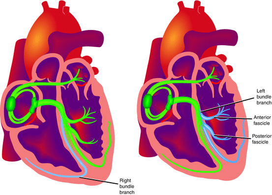
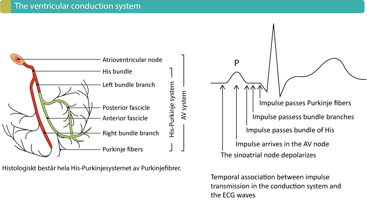
Sinus rythm with marked sinus arythmia incomplete right bundle branch block Borderline ECG ECG results 79 pbm, Pr interval 152 ms, Qrs duration 100 ms,QT/QTc 352/403 ms, p r t axes 21 17 Is this something I should be worried about?The main effects of ventricular preexcitation by an accessory pathway during sinus rhythm are seen in the first part of the QRS complex on the surface ECG The degree of preexcitation seen depends on the degree of its fusion with conduction down the HisPurkinje system1 In patients with aRight bundle branch block (RBBB) Right bundle branch block (RBBB) is due to an anatomical or functional dysfunction in the right bundle branch, such that the electrical impulse is blocked Refer to Figure 1 for an overview of the components of the ventricular conduction system, including the right bundle branch
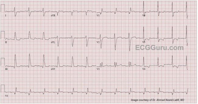


Right Bundle Branch Block Ecg Guru Instructor Resources
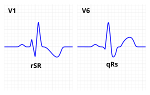


Right Bundle Branch Block Wikipedia
Avb 2nd degree, mobitz i (wenckebach) av block 2nd degree, mobitz ii (hay) avb 2nd degree, 21 conduction;Apr 15, 05 · Fascicular block refers to electrocardiographic (ECG) evidence of impaired conduction below the AV node in the right bundle branch or in one or both fascicles of the left bundle branchJan 08, 21 · Atrioventricular (AV) conduction is evaluated by assessing the relationship between the P waves and QRS complexes Normally, there is a P wave that precedes each QRS complex by a fixed PR interval of 1 to 0 milliseconds AV block represents a delay or disturbance in the transmission of an impulse from the atria to the ventricles This can be due to an anatomical or


The 12 Rhythms Of Christmas First Degree Av Block Ems 12 Lead


Case B5 Second Degree Av Block Mobitz Type I Or Wenckebach Block St Emlyn S Ecg Library St Emlyn S
Jun 13, 16 · Right bundlebranch block (RBBB) may be present in healthy individuals with no apparent underlying heart disease, but more commonly occurs in the presence of coronary artery disease (the most common cause) RBBB may be temporary or chronic Occasionally, RBBB may appear only when the heart rate exceeds a certain critical level (raterelated BBB)Feb 06, 18 · Her baseline ECG (see Figure 1) showed sinus rhythm (SR), heart rate 87/min, left ventricular hypertrophy with repolarisation abnormalities and normal PR, QRS and QTc intervals Her baseline ECG transformed into sinus tachycardia with left bundle branch block (LBBB) pattern (see Figur e 2) Atrioventricular (AV) pacemaker was inserted forPseudo right bundle branch block;


The 12 Rhythms Of Christmas First Degree Av Block Ems 12 Lead
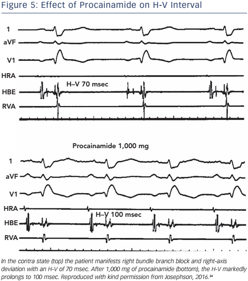


Electrophysiological Testing For The Investigation Of Bradycardias Aer Journal
2 days ago · The PR interval is normally 01 seconds or 1 to 0 milliseconds A PR interval consistently longer than 0 seconds (greater than 5 small boxes) indicates a 1st degree AV blockSinus bradycardia ECG, causes & management Definition of sinus bradycardia Sinus bradycardia fulfills the criteria for sinus rhythm but the heart rate is slower than 50 beats per minute ECG criteria follows Regular rhythm with ventricular rate slower than 50 beats per minute Pwaves with constant morphology preceding every QRS complexAnd ii) there is a hint of ST elevation with T wave inversion in lead III — suggesting recent inferior infarction (and Mobitz I is much more common in association with inferior infarction) Other STT wave changes on this
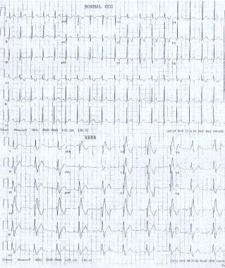


Pediatric Right Bundle Branch Block Background Pathophysiology Etiology


18 Acc Aha Hrs Guideline On The Evaluation And Management Of Patients With Bradycardia And Cardiac Conduction Delay Heart Rhythm
Posted by Shirley on May 11, 1999 at My husband was recently diagnosed with Sinus Bradycardia Right Bundle Branch Block He is 34 years old His heart rate was 49bpm His blood pressure was 140/80 His physician said it was nothing for my husband to worry about but I am looking for a 2nd opinionAvb 2nd degree, "high grade av block" avb 3rd degree (complete heart block) avnrt (av nodal reentry tachycardia) avrt (av reentry tachycardia) brash syndrome;Bundle Branch Block •Problem with branches of the ventricular conduction system •This is below the area of a "Heart Block" Heart block may be delayed or blocked in the ___ •AV node •Below the AV node •Bundle of HIS Causes of temporary heart block
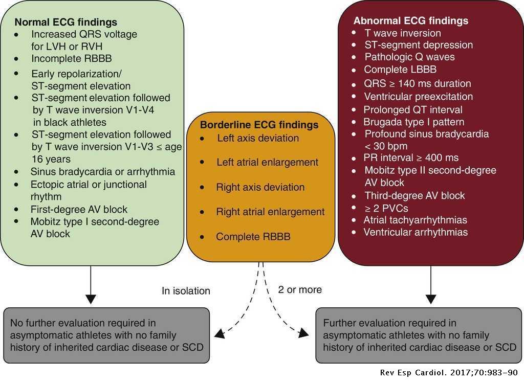


Comments On The New International Criteria For Electrocardiographic Interpretation In Athletes Revista Espanola De Cardiologia


Cardiovascular Medicine Bradyarrhythmias
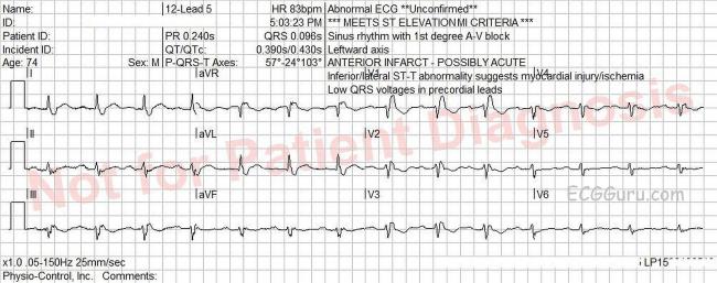
Apr 07, 21 · Firstdegree atrioventricular (AV) block is a condition of abnormally slow conduction through the AV node It is defined by ECG changes that include a PR interval of greater than 0 without disruption of atrial to ventricular conduction This condition is generally asymptomatic and discovered only on routine ECGMar , 21 · Sinus Rhythm with 2nd or 3rd degree AV Blocks Atria are depolarizated by sinus node, but some (2nd degree AV block) or all (complete AV Block) stimuli are not conducted to the ventricles In 2nd degree AV block not all sinus P waves are followed by QRS complexes because there is an intermittent failure of the AV conductionThe criteria to diagnose a right bundle branch block on the electrocardiogram The heart rhythm must originate above the ventricles (ie, sinoatrial node, atria or atrioventricular node) to activate the conduction system at the correct point The QRS duration must be more than 100 ms (incomplete block) or more than 1 ms (complete block)


Bifascicular Blocks What You Need To Know Ecg Medical Training


Bifascicular Blocks What You Need To Know Ecg Medical Training
Please can somone clarify further many thanksRight And Left Bundle Branches First Degree Av Block Third Degree Heart Block Second Degree Heart Block Coffee And Tea TERMS IN THIS SET (36) The licensed practical nurse is coassigned with a registered nurse in the care of a client admitted to the cardiac unit with chest painAll P waves conduct to the ventricles 2nd Degree AV Block
:max_bytes(150000):strip_icc()/iStock-616893490-59270be03df78cbe7eef363d.jpg)


When Is A Pacemaker Needed For Heart Block


File Rbbb With First Degree Av Block Jpg Wikimedia Commons
Feb 03, 13 · This year old man has a sinus rhythm that is around 100 bpm, and his QRS is widened at 148 ms (148 sec) Leads I and V6 are positive, and Lead V1 is negative, meeting the criteria for left bundle branch block There is a left axis deviation, which is common with LBBB, although it is not always this pronounced, indicating that there isRight bundle branch block comes from a problem with the heart's ability to conduct electrical signals It usually doesn't cause symptoms unless you have some other heart condition Your heart has 4 chambers The 2 upper chambers are called atria The 2 lower chambers are called ventricles In a healthy heart, the electrical signal for yourJan 06, · Firstdegree atrioventricular (AV) block, or firstdegree heart block, is defined as prolongation of the PR interval on an electrocardiogram (ECG) to more than 0 msec The PR interval of the surface ECG is measured from the onset of atrial depolarization (P wave) to the beginning of ventricular depolarization (QRS complex)


Left Bundle Branch Block Ecg 4 Learntheheart Com
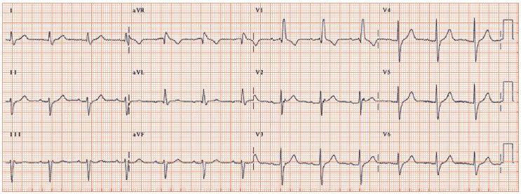


Right Bundle Branch Block Thoracic Key
Incompleterightbundlebranchblock & Sinusbradycardia Symptom Checker Possible causes include Cardiomyopathy Check the full list of possible causes and conditions now!Jan 14, · An escape rhythm, while lifesaving, is often unreliable for prolonged periods of time In general, the higher the degree of heart block, the more likely the need for a pacemaker Pacemakers are almost always required with thirddegree block, often with seconddegree block, but only rarely with the firstdegree blockJun , 18 · The bundle of His is an area of the heart that conducts impulses to the left and right ventricles Left bundle branch block (LBBB) is a blockage of


Lbbb With First Degree Av Block


The 12 Rhythms Of Christmas First Degree Av Block Ems 12 Lead
First degree AV block does not generally cause any symptoms, but may progress to more severe forms of heart block such as second and thirddegree atrioventricular block It is diagnosed using an electrocardiogram, and is defined as a PR interval greater than 0 millisecondsOne narrow (yellow highlight) and one wide with a right bundle branch block configuration (red highlight) This is a bidirectional rhythm (or tachycardia if greater than 100Jun , 18 · The bundle of His is an area of the heart that conducts impulses to the left and right ventricles Right bundle branch block (RBBB) is a blockage of electrical impulses to the heart's right
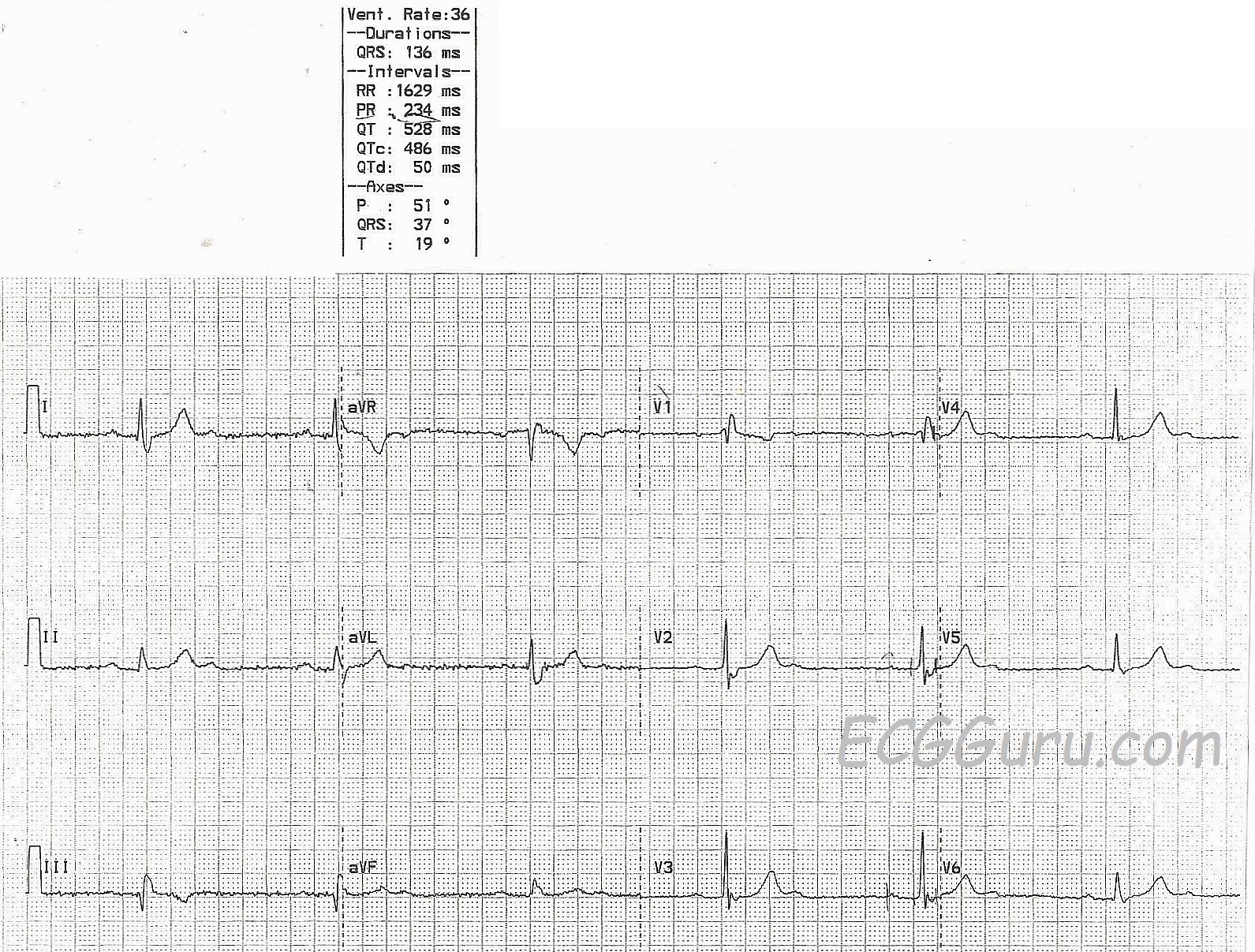


Second Degree Av Block With 2 1 Conduction And Right Bundle Branch Block Ecg Guru Instructor Resources


Electrocardiogram Demonstrating Normal Sinus Rhythm With First Degree Download Scientific Diagram
Possible sites of AV block AV node (most common) His bundle (uncommon) Bundle branch and fascicular divisions (in presence of already existing complete bundle branch block) 1st Degree AV Block PR interval > 0 sec;Bifascicular block is a conduction delay or "block" below the atrioventricular (AV) node in two of the three fascicles (the right bundle branch and left anterior and left posterior fascicles of the left bundle branch) Right bundle branch block (RBBB) is typically combined with either left anterior fascicular block (LAFB) or left posteriorTalk to our Chatbot to narrow down your search


Phase 4 Block Of The Right Bundle Branch Suggesting His Purkinje System Involvement In Lyme Carditis Heartrhythm Case Reports


A 46 Year Old Man With Burning Epigastric Pain Of Several Hours Duration Page 2 Of 2 Journal Of Urgent Care Medicine
Persistent seconddegree AV block Mobitz type II and/or thirddegree block Right Bundle Branch Block diagnosed by presence of RSR' waves in V1 and/or V2 and QRS complex wider than 012 seconds (3 small blocks)Rhythm analysis indicates normal sinus rhythm (NSR) with a 1st degree heart block This encounter displays a P wave of over 0 ms or one square away from the QRS, making it 1st degree heart block This condition represents a delay between atrial and ventricular conduction It is not considered dangerous, but can escalate to higher level blocksA, The electrocardiogram shows normal sinus rhythm Two P waves conduct to the ventricle, followed by the third P wave (high frequency deformity in the T wave) failing to conduct B, Surface recordings (leads I, II, and V 1) show normal sinus rhythm with Mobitz type 1 seconddegree atrioventricular (AV) block and right bundlebranch block


Ecg Sinus Rhythm 1st Degree Av Block Left Bundle Branch Block Download Scientific Diagram


Baseline Left Bundle Branch Block With Right Bundle Branch Escape Complexes In A Patient With Coronary Artery Disease Presents Like An Alternating Bundle Branch Block A Case Report Cases Journal
Firstdegree heart block, or firstdegree AV block, is when the electrical impulse moves through the AV node more slowly than normal The time it takes for the impulse to get from the atria to theFirstdegree AV block is an abnormal delay in conduction through the AV node This type of AV block is a disturbance in the conduction between the normal sinus impulse and its eventual ventricular response This manifests as a prolonged PR interval on ECG Meanwhile, the heart is maintained in sinus rhythm, with a normal QRS configuration


First Degree Av Block Ekg Ecg Interpretation Youtube
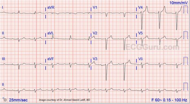


Wide Qrs Complex With First Degree Av Block Ecg Guru Instructor Resources


1
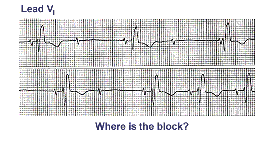


Ecg Learning Center An Introduction To Clinical Electrocardiography


Bradyarrhythmias And Conduction Blocks Revista Espanola De Cardiologia


Q Tbn And9gctean9xupbblxw2uu310w86eanw8kuua6jymq290fuhxxv8y02o Usqp Cau


Heart Rhythm Guide
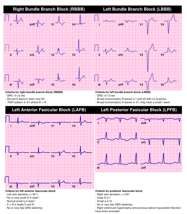


Bradycardia Textbook Of Cardiology


Surgical Complete Heart Block Alternating With Sinus Rhythm And Right Bundle Branch Block With Left Anterior Hemiblock Chest
/right-bundle-branch-block-rbbb-1745785-FINAL-35a6f733fab44ef08240c21a675c12da.png)


Bundle Branch Block Overview And More


Atrioventricular Block Cardiovascular Disorders Merck Manuals Professional Edition


The 12 Rhythms Of Christmas First Degree Av Block Ems 12 Lead


Bifascicular Blocks What You Need To Know Ecg Medical Training


Bundle Branch Block Animation Youtube


B24 Right Bundle Branch Block With The Wenckebach Phenomenon In Acute Myocardial Infarction St Emlyn S


Right Bundle Branch Block Disease Malacards Research Articles Drugs Genes Clinical Trials
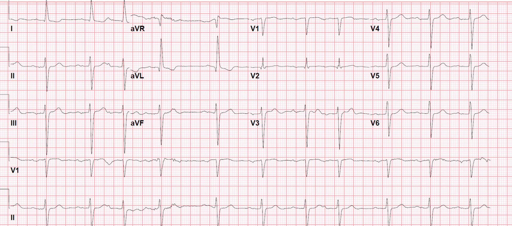


Cureus Left Bundle Branch Block A Reversible Pernicious Effect Of Lacosamide


Understanding Atrioventricular Blocks Careercert
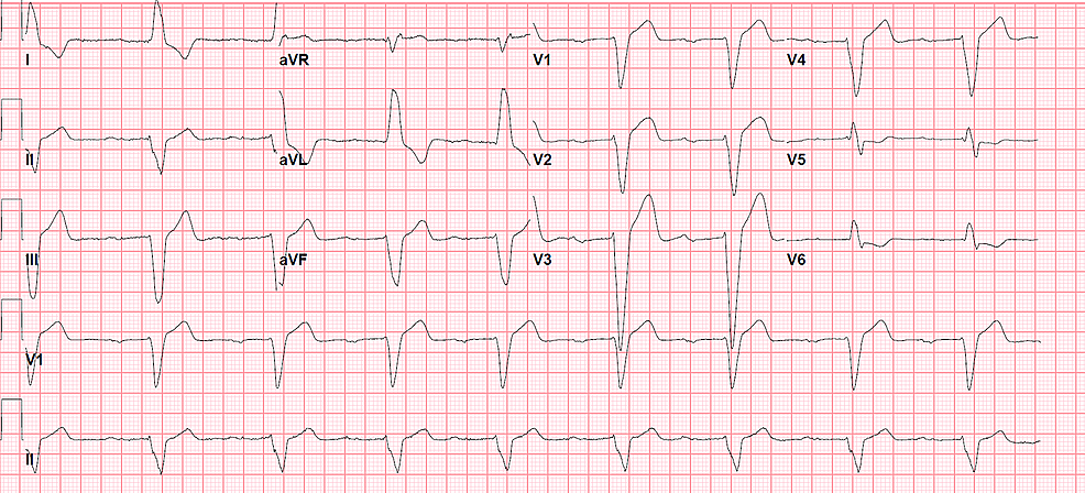


Cureus Left Bundle Branch Block A Reversible Pernicious Effect Of Lacosamide


1


Bundle Branch Block Causes Symptoms Diagnosis Treatment
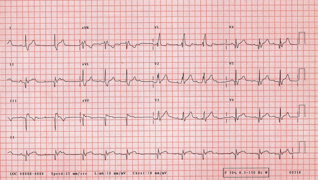


B24 Right Bundle Branch Block With The Wenckebach Phenomenon In Acute Myocardial Infarction St Emlyn S


The 12 Rhythms Of Christmas First Degree Av Block Ems 12 Lead


19 The Basis Of Ecg Diagnosis
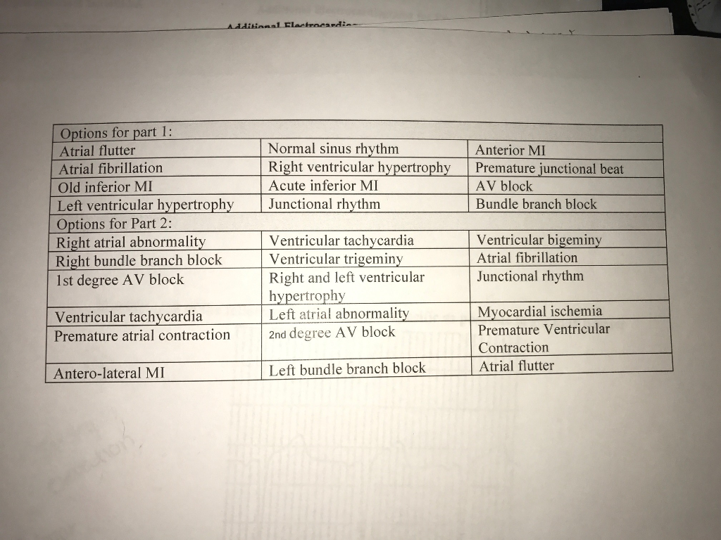


Ekq Abnormalities Questions Please Help If You U Chegg Com
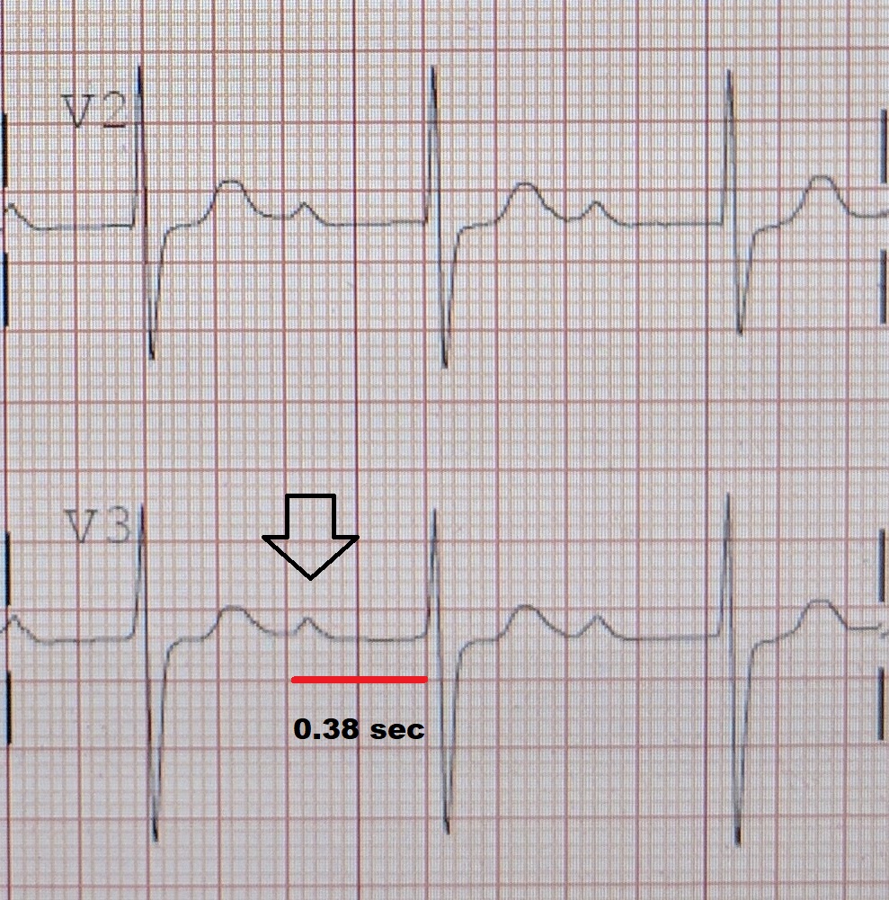


First Degree Atrioventricular Block Wikipedia


Heart Blocks Explained First Second Third Degree And Bundle Branch On Ecg Youtube


3rd Degree Av Block Ecg 1 Learntheheart Com


Arrhythmias


Bifascicular Blocks What You Need To Know Ecg Medical Training
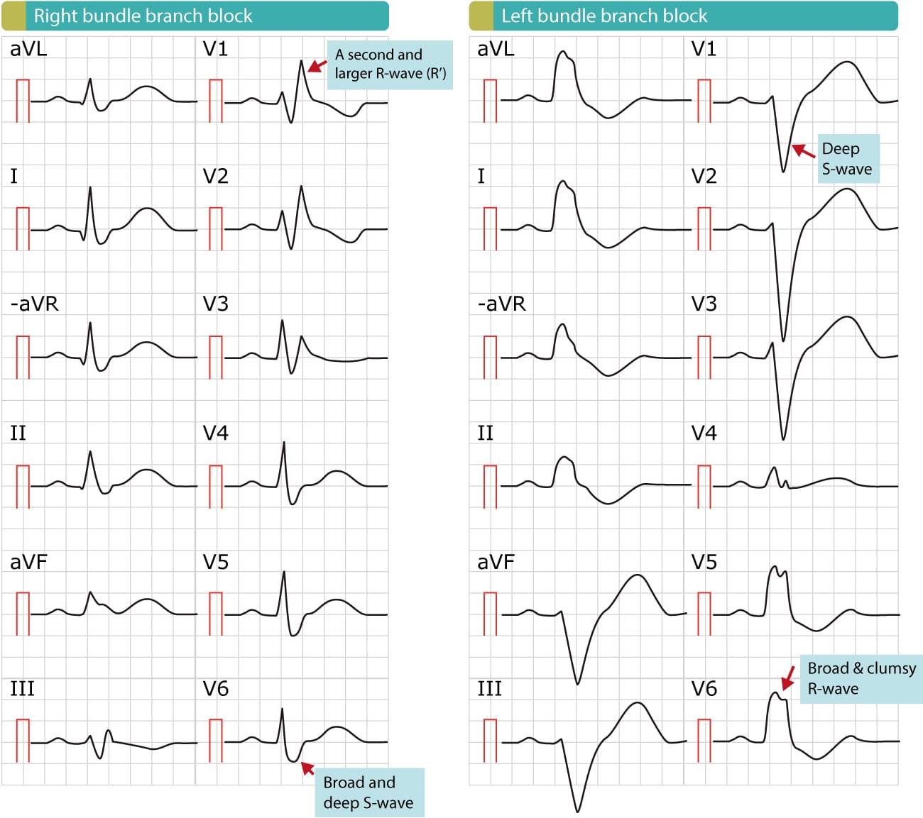


Right Bundle Branch Block Rbbb Ecg Criteria Definitions Causes Treatment Ecg Echo


Cardiovascular Medicine Bradyarrhythmias


Lbbb With First Degree Av Block


1


Ekg Learn The 4 Types Of Atrioventricular Blocks Leveluprn


Ecg Learning Center An Introduction To Clinical Electrocardiography


Indications And Recommendations For Pacemaker Therapy American Family Physician


Bundle Branch Block Wikipedia


Alternating Bundle Branch Block Circulation


Ecg Learning Center An Introduction To Clinical Electrocardiography


Intraventricular Conduction Defect Trifascicular Block Basic And Bedside Electrocardiography 1st Edition 09
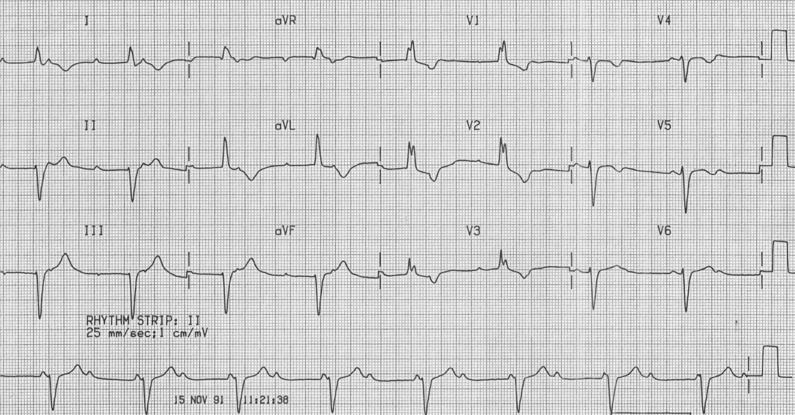


Trifascicular Block Litfl Ecg Library Diagnosis


Evaluation And Treatment Of Av Block And Intraventricular Conduction Disturbances The Cardiology Advisor


Evaluation And Treatment Of Av Block And Intraventricular Conduction Disturbances The Cardiology Advisor
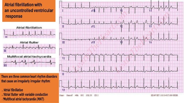


Ecg Quiz


Ekg Interpretation


Progressive First Degree Atrioventricular Block As A Warning Sign For Perioperative Bradyarrhythmia Journal Of Cardiothoracic And Vascular Anesthesia


Dr Smith S Ecg Blog Intermittent Third Degree Heart Block Due To Stuttering Inferior Stemi
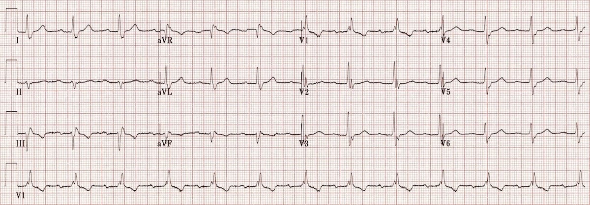


Trifascicular Block Litfl Ecg Library Diagnosis


Atrioventricular Block Amboss
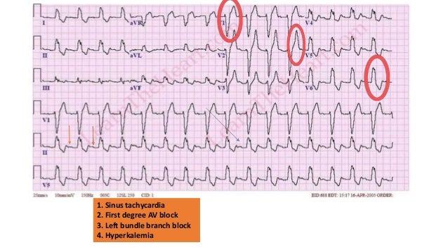


Ecg Quiz


First Degree Av Block Av Block I Av Block 1 Ecg Echo


Pediatric Third Degree Acquired Atrioventricular Block Background Classification Of Atrioventricular Blocks Pathophysiology


Conduction Blocks And Cardiac Pacing Thoracic Key


Ecg Learning Center An Introduction To Clinical Electrocardiography


Ecg Sinus Rhythm bpm Qrs Axis 10 First Degree Atrioventricular Download Scientific Diagram


Heart Block Wikipedia


Third Degree Atrioventricular Block Atrioventricular Node Atrium Sinoatrial Node Bundle Branch Block Qrs Png Pngegg


Overview Of Atrioventricular Av Blocks Ecg Echo


Heart Block Causes Symptoms Types Diagnosis And Heart Block Treatment


Right Bundle Branch Block With Machine Interpretation Error Ecg Guru Instructor Resources
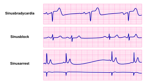


Bradycardia Textbook Of Cardiology


Ekg S Internal Medicine University Of Nebraska Medical Center
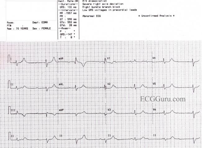


Third Degree Av Block And Junctional Escape Rhythm With Right Bundle Branch Block And Prolonged Qtc Interval Ecg Guru Instructor Resources


A During Sinus Rhythm Heart Rate Of 110 Min Right Bundle Branch Download Scientific Diagram


A 46 Year Old Man With Burning Epigastric Pain Of Several Hours Duration Page 2 Of 2 Journal Of Urgent Care Medicine


Dr Smith S Ecg Blog Intermittent Third Degree Heart Block Due To Stuttering Inferior Stemi
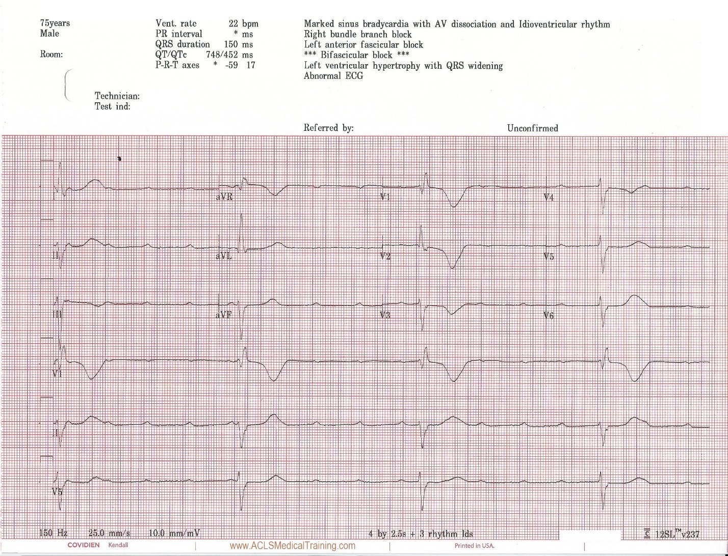


Unstable Bradycardia Resolves Following Atropine And Attempted Transcutaneous Pacing Tcp Acls Medical Training


Twelve Lead Electrocardiogram Sinus Rhythm Right Bundle Branch Block Download Scientific Diagram


Anaesthesia Uk Conduction Abnormalities


First Degree Atrioventricular Block An Overview Sciencedirect Topics


Right Bundle Branch Block Wikipedia


How To Recognize Treat Heart Block Jems


How To Recognize Treat Heart Block Jems


Bundle Branch Block Wikipedia


View Image


コメント
コメントを投稿